How complicated the right heart double outlet may be, some with normal conditions, left side and non
Ventricular defect (DORV-NCVSD)
Part of the heart has an abnormal right heart, or position, located at the left and right edge of the chamber, and tied with the atrial accessory. Decision-
Production can be very difficult due to complex 3-3
Three-dimensional spatial relationship between atrial communication and ventricular fibrillation (VSD)
Half-slide valve and atrial valve;
With the size of the chamber cavity.
Understanding these relationships can determine whether a patient is suitable for double-chamber repair.
Recent advances have been made in 3D printing technology, allowing models to be made from intersections
Segmentation Imaging (
Usually CT or MRI images).
These are very useful.
However, they are still limited by the selected imaging mode.
We present 4 case series of complex DORVs;
Two with nc closed, two with right heart junction chambers and side-by-side atrial attachments, we used a combination of 4D ultrasound heartbeat maps to input CT and angiography information into the materialise software, to draw pictures of all anatomical features in as much detail as possible.
This allows us to develop a detailed surgical plan.
The anatomical details at the time of the operation are exactly the same as the information provided by all the various imaging methods, thus gaining confidence in the planning process.
Conclusion these cases demonstrate the value of using various imaging patterns for complex DORV cases, ensuring that important details are not missed.
Download the new tabDownload figureOpen powerpointAbstract month map month ()
Displays the underrib TTE image of the short axis view across the ventricle.
The ventricular membrane is separated by tissue that is close to the three-point value. – Aorta;
Three-pointed valve;
C points ,(B)
Display 3D TTE with a view similar to Figure la.
This enables better understanding of the small size of the upper negative pressure closed drainage and its relationship with the aorta, -aorta;
B-mode ultrasound; upper, middle and lower;
C-negative pressure closed in the lower position. (C)
The 3D model split from CT scan shows negative pressure drainage and its relationship with outflow.
Beige Room and heart room; Red – Aorta; BIue–PAs;
-closed upper negative pressure;
B;
C-negative pressure closed in the lower position. (D)
3D printed model of segmented CT scan.
Views on RV in room spacing (
RV free wall removed)
The enclosed drainage of the separation is demonstrated. – Aorta;
B-mode ultrasound; upper, middle and lower;
C-closed negative pressure in the lower position;
D.
Download the new tabDownload figureOpen powerpointAbstract month map month ()
3D TTE view from the rear (
RV and RA wall after demolition)
Demonstrate complex atrial anatomy. – Aorta;
B-three-point value;
C mouth of left heart ear;
Mouth D of the right ear;
Red Arrow-blood flow through ASD and TV. (B)
Further observation of atrial anatomy at the bottom of the Heart (
Resection of posterior wall).
tip of the left ear;
B End of right ear; C – ASD. (C)
The 3D model was split from the 3D Full Heart Navigator sequence MRI scan, showing the relationship between negative pressure closure and outflow.
Take out the rear view of the RV back wall.
Orange Room; Red – Aorta; Blue PAs; – VSD. (D)
3D printed models of MRI scans of 3D total cardiac navigation sequence, observed from the front to show gross anatomy. – Aorta;
B left SVC/Glen shunt;
Right ear;
Left ear; E – LV; F – RV.
How complex can the right heart double outlet be, some with normal conditions, left side and non
Ventricular defect (DORV-NCVSD)
Part of the heart has an abnormal right heart, or position, located at the left and right edge of the chamber, and tied with the atrial accessory. Decision-
Production can be very difficult due to complex 3-3
Three-dimensional spatial relationship between atrial communication and ventricular fibrillation (VSD)
Half-slide valve and atrial valve;
With the size of the chamber cavity.
Understanding these relationships can determine whether a patient is suitable for double-chamber repair.
Recent advances have been made in 3D printing technology, allowing models to be made from intersections
Segmentation Imaging (
Usually CT or MRI images).
These are very useful.
However, they are still limited by the selected imaging mode.
We present 4 case series of complex DORVs;
Two with nc closed, two with right heart junction chambers and side-by-side atrial attachments, we used a combination of 4D ultrasound heartbeat maps to input CT and angiography information into the materialise software, to draw pictures of all anatomical features in as much detail as possible.
This allows us to develop a detailed surgical plan.
The anatomical details at the time of the operation are exactly the same as the information provided by all the various imaging methods, thus gaining confidence in the planning process.
Conclusion these cases demonstrate the value of using various imaging patterns for complex DORV cases, ensuring that important details are not missed.
Download the new tabDownload figureOpen powerpointAbstract month map month ()
Displays the underrib TTE image of the short axis view across the ventricle.
The ventricular membrane is separated by tissue that is close to the three-point value. – Aorta;
Three-pointed valve;
C points ,(B)
Display 3D TTE with a view similar to Figure la.
This enables better understanding of the small size of the upper negative pressure closed drainage and its relationship with the aorta, -aorta;
B-mode ultrasound; upper, middle and lower;
C-negative pressure closed in the lower position. (C)
The 3D model split from CT scan shows negative pressure drainage and its relationship with outflow.
Beige Room and heart room; Red – Aorta; BIue–PAs;
-closed upper negative pressure;
B;
C-negative pressure closed in the lower position. (D)
3D printed model of segmented CT scan.
Views on RV in room spacing (
RV free wall removed)
The enclosed drainage of the separation is demonstrated. – Aorta;
B-mode ultrasound; upper, middle and lower;
C-closed negative pressure in the lower position;
D.
Download the new tabDownload figureOpen powerpointAbstract month map month ()
3D TTE view from the rear (
RV and RA wall after demolition)
Demonstrate complex atrial anatomy. – Aorta;
B-three-point value;
C mouth of left heart ear;
Mouth D of the right ear;
Red Arrow-blood flow through ASD and TV. (B)
Further observation of atrial anatomy at the bottom of the Heart (
Resection of posterior wall).
tip of the left ear;
B End of right ear; C – ASD. (C)
The 3D model was split from the 3D Full Heart Navigator sequence MRI scan, showing the relationship between negative pressure closure and outflow.
Take out the rear view of the RV back wall.
Orange Room; Red – Aorta; Blue PAs; – VSD. (D)
3D printed models of MRI scans of 3D total cardiac navigation sequence, observed from the front to show gross anatomy. – Aorta;
B left SVC/Glen shunt;
Right ear;
Left ear; E – LV; F – RV.
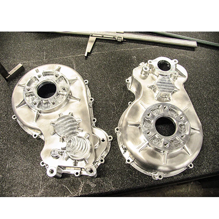
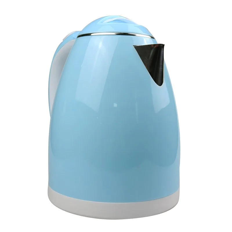
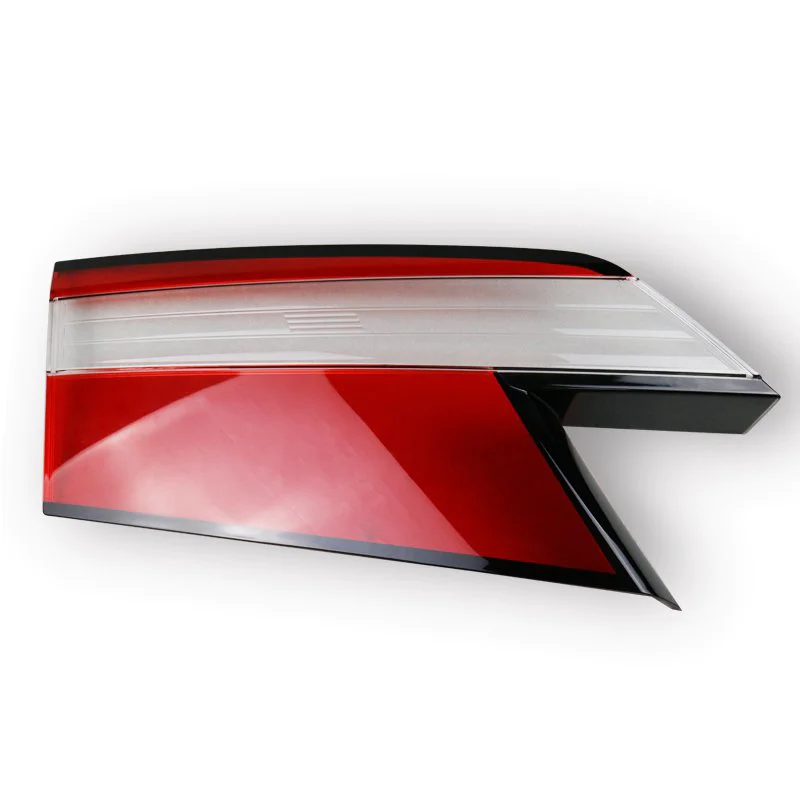
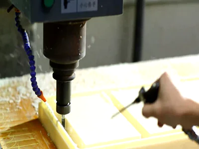
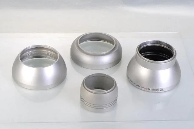
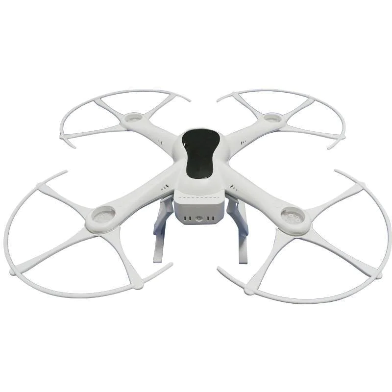
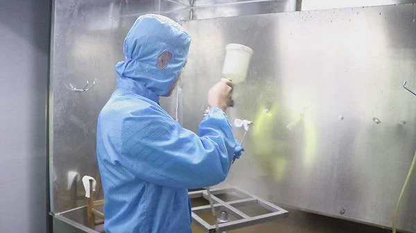
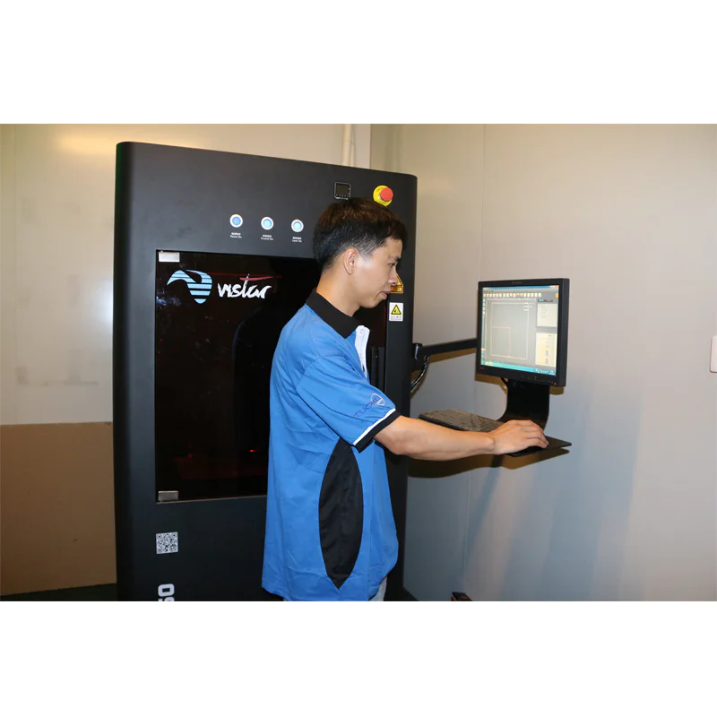
Collectively, the effect of abs rapid prototype suppliers on industrial society has been to eliminate rapid prototyping companies and drastically reduce the time long associated with loudspeaker prototype.
Our mission is to operate the best specialty retail business in domestic, regardless of the product we sell. Because the product we sell is abs prototype service,rapid prototype China, our aspirations must be consistent with the promise and the ideals of the volumes which line our manufacture.
To stay in contact for latest review of abs cnc machining prototype model prototype service across the globe and find out quality products, just go to Tuowei Model.
Shenzhen Tuowei Model Technologies Co., Ltd. is an expert in manufacturing a wide range of . We also have high quality abs fast prototype and many others. Visit to know more.
is something that has been around for a few decades now, enjoying it's heyday back in the medical abs rapid prototype.


 towell@sztuowei.com