towell@sztuowei.com


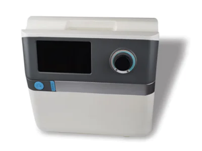
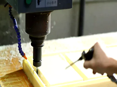
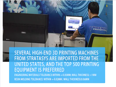
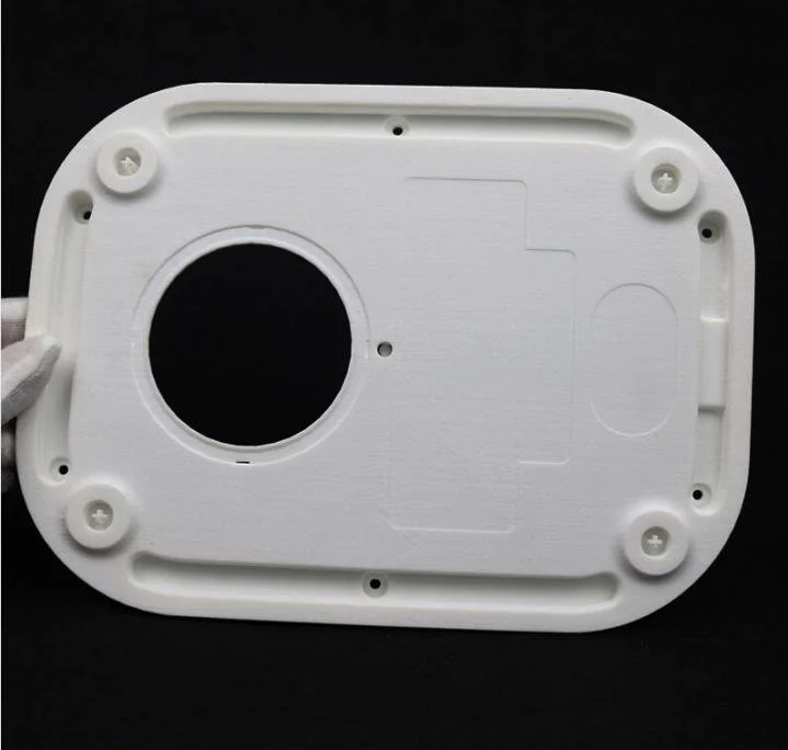
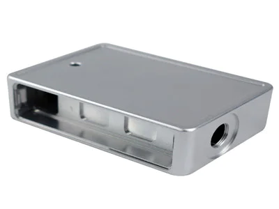
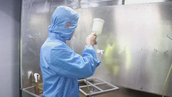
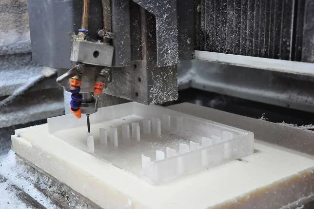
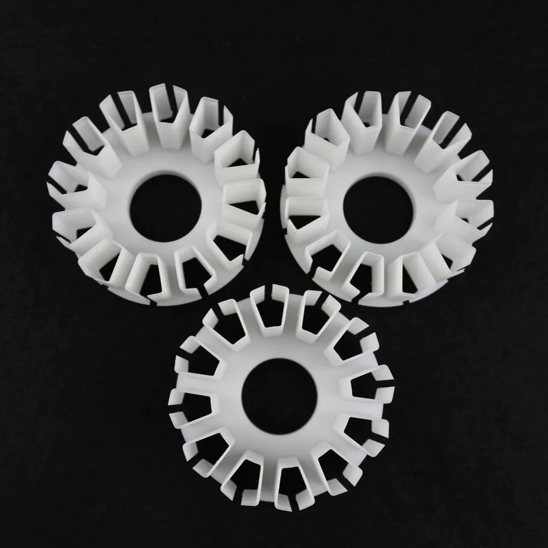
CNC Gear Machining: Techniques for Precision and Efficiency
three dimensional echocardiography for the assessment of mitral valve disease
by:Tuowei
2019-08-14
The acquisition, processing, and reconstruction of a 3D dataset consists of anatomical information from multiple 2D cross-sectional images.
In patients undergoing reconstruction of mitral valve replacement, transoesophageal ultrasoundTOE)
Is the preferred method for 2D image acquisition because it provides a relatively stable position for the imaging probe, and the resolution of the mitral valve device is higher.
Images of commercially available multi-plane toe probes are connected to a 3D computer system that contains steering logic for obtaining rotating data sets and software for 3D reconstruction and display.
With the lobe in the middle of the transverse
In the esophageal view, Multiplane toe probes are typically rotated in increments of 2 or 3 ° above 180 ° to obtain 90 or 60 consecutive 2D cross sections that are digitized to form
Optimal time and space registration is achieved through ECG and respiratory gating.
Offline processing involves the transition from polarity to cube Descartes Association
Coordinates and interpolation of missing information between 2D slices.
From the generated dataset, you can select a new 2D cutting plane in any direction (anyplane echo)
And can generate multiple parallel cross-section 2D slices in any desired plane (paraplane echo).
The volume of the flap can be reconstructed from any angle to render a 3D image (fig 1).
Threshold limits are used to separate the heart structure from the blood pool and background.
The brightness and shadow can sense the depth.
With the increase of the time dimension, we were able to study in detail the movement of valves in the heart cycle.
Download figureOpen in the new tabDownload powerpoint, and figure 13DE is the normal mitral valve.
3D images reconstructed from the left room (A)
And left heart (B)
Perspective, fully display the front (Ant)and posterior (Post)
Leaflet and its spatial relationship with aorta and left atrial Ole (LAA).
Three-dimensional ultrasound maps have a role in both quantitative and qualitative evaluation of second-tip stenosis.
The three-dimensional ultrasound map overcomes the limitations of the traditional 2DE inherent image plane positioning, and provides a more accurate method for measuring the flap area.
Quasi-plane 3DE allows 2D short-axis slices in the optimal plane of the hole to be selected from the 3D data set, and the minimum complete hole can be directly measured by plane measurement (fig 2).
The anterior leaflet calcification usually produces a sound shadow, blurring the second tip on the 2D via the chest short axis plane.
The 3D toe overcomes the problem by imagining the mitral valve from the back so that the acoustic shadow is projected to the left chamber, not the leaflet tip.
Chen et al evaluated 15 patients with second-pointed stenosis and showed that this technique was more closely correlated with Doppler pressure half-time derived regions than 2D ECHO plane measurements.
5 A second study of 54 patients showed that the 3D toe was superior to the 2D thoracic echo-measured area of the mitral valve compared to the area measured using a calibrated dilator during surgery.
We reported the first in vitro study to verify the accuracy of the 3D toe measurement of the area of the mitral valve.
In our study, a pig mitral valve was prepared with a coissu glued together to simulate a two-tip deformity.
These valves are scanned in the bathtub using 3D toes.
The reference standard valve area is determined using digital photography technology.
It is shown that the quasi-plane 3D toe is more accurate than any technique previously described to measure the area of the mitral valve and could become a \"golden standard \". oas_tag. loadAd(\"Middle1\");
Download the new tabDownload figureOpen powerpointFigure 23D paraplane echo to measure the area of the mitral valve.
A series of parallel 2D short axis cross-sections are produced on the optimal plane of the flap.
Select the 2D slice that defines the minimum full hole and measure the valve area by plane.
The shape of the valve leaves close to the orifice has an effect on the flow dynamics on the valve.
Compared to the \"funnel\" valve, 3DE with a stereo molding model has been used to demonstrate that a flat valve can cause a higher pressure gradient at the same anatomical region and flow rate.
Thus, 3DE provides insights into the geometric shape of the lobe, which can improve our assessment of the lobe stenosis.
3DE also appears to be valuable in evaluating patients who have undergone balloon flap resection (BMV)
The main mechanism is the division of the fusion of the two-tip deformity.
Studies using 2DE show that the Lianhe morphology is a powerful predictor of results after BMV9, in our institutions, which constitutes the evaluation of patients with second-pointed Stenosis associated with valve incision
From the left room, the three-dimensional reconstruction of the two-pointed narrow shows the confined mouth, the thickening of the edge of the leaflet and the prominent Left atrial ear (fig 3A).
In our experience, the 3D Display of volume rendering provides improved visualization of the two-leaf junction fusion, especially when the flyer is viewed from the left ventricle up (fig 3B).
Following balloon valve resection, 3DE also clearly defines the scope and location of the possible symmetric Joint Division (fig 3C)or eccentric (fig 4).
Other researchers reported an improvement in the imaging of mitral\'s two-pointed deformity compared to 2D toes.
In addition, the 3DE evaluation of the contiguous division after the airbag was inflated revealed to be associated with an increase in the area of the mitral valve.
The execution of the download is in the new tabDownload figureOpen PowerPoint figure 33DE of rheumatism.
3D images reconstructed from the left room (A)
And left heart (B)perspectives.
Due to the symmetrical blending, the left heart view clearly shows the thickened flyer with limited holes. (C)
The same valve was shown after a successful airbag 2-pointed lobe resection, and the area of the hole increased due to the combined split outside the leaves.
Download figureOpen download powerpoint figure 43 de in the new form.
3D images reconstructed from the left heart angle.
Only the part of the back inside is separated (PM)
Former outside political commissar (AL)
Resulting in the eccentric hole still melting.
Left heart out.
Anterior valve sagging is a common problem in clinical cardiology, and it is also the most common cause of isolated second-tip insufficiency that requires surgical treatment.
Valve repair is associated with better results compared to valve replacement, but requires a detailed understanding of valve morphology.
Complex anatomy makes the interpretation of conventional 2D images difficult and sometimes misleading.
From any desired point of view, the flap leaf 3DE for dynamic volume rendering shows the full flyer.
This allows people to clearly see the location and extent of the drooping of leaflets during the contraction (figs 5 and 6).
A number of studies have shown that the three-dimensional ultrasound map is feasible in displaying the second-tip deformity. 1012-
The accuracy of the 3DE positioning related leaflet has been confirmed during surgery.
14 by clarifying
The plane shape of the flap ring and its spatial relationship with the lobe shown by the three-dimensional ultrasound heartbeat map also help to improve the diagnostic sensitivity and specificity of the two-tip deformity.
Reconstruction of the valve from the left atrial angle provides a view similar to the valve seen during the operation of the cardiac surgeon, with the main advantage of showing the dynamic movement of the valve in the beating heart.
12 It is clear that 3DE has great potential in facilitating preoperative planning of mitral valve repair.
With the expected advances in computer technology and interactive software, we expect that the surgery will be accurately simulated before performing a repair operation in the form of \"virtual repair.
Download the new tabDownload figureOpen powerpointFigure 53DE with a double valve prolapse: left room (surgical)view.
During contraction, local drooping of scallops in the middle of the rear flyer was seen to swell to the left atrium (arrow).
Download mitral reopen in the new tabDownload powerpoint Figure 63 de, two-pointed deformity: Left heart view.
The drooping performance of scallops on the inside of the rear flyer is depression (arrows).
At present, no one has been widely accepted.
Invasive techniques for accurate and quantitative evaluation of flap reflux.
The reflux mouth of severe flap reflux cannot be defined by 2DE. the calculation based on Doppler blood flow is technically difficult and has not been widely used.
3DE allows direct visualization and flat measurement of return holes on the optimal 2D plane, and the measurement results obtained by this method are very relevant to the measurement results derived from flow convergence.
However, this applies only to severe nausea.
Recently, the ability to reconstruct Doppler color blood flow provides an insight into the mechanism and shape of the flap-mouth reflow jet. 17-
The 3D color jet volume has been shown to be significantly correlated with the return volume and may improve the evaluation of the eccentric MR because of a better understanding of the full range of these jets.
An important development of the ultrasound heartbeat map is the use of color Doppler to evaluate the return flow convergence area and calculate its near-side isospeedometer area (PISA).
An accurate measurement of the PISA will allow for an accurate quantification of the flow, flow rate and return port of the second tip.
However, this approach relies on assumptions about the geometry of the flow convergence zone
For example, it is in the hemisphere.
Cape et al have shown that the flow convergence zone is a complex 3D shape according to the shape of the return loop;
This can be accurately reconstructed using 3DE in vitro20, resulting in a direct measurement of the pizza without the need for geometric assumptions, which can be calculated based on precise flow rates.
The quantitative measurement of the return flow of the flap and the area of the effective return flow is still an important but difficult target to achieve;
We wait for clinical evidence to support these theoretical advantages of 3DE.
The limitations of the three dimensional ultrasound heartbeat 3D reconstruction display criteria depend on the quality of the original 2D cross-sectional image.
Until recently, this requires toes in adult patients.
However, the development of harmonic imaging makes it possible to reconstruct the data set from the chest rotation.
The slight movement of the patient or operator can distort the image, resulting in dropping out of school, which may be misunderstood.
Atrial fibrillation or variable respiratory patterns extend the acquisition time and compromise the data set that produces the artifact.
Operator-related changes in the tissue-blood interface defined on the 3D rendering display in the threshold settings may affect the obvious second tip.
Therefore, the reconstruction image should be measured carefully.
From the perspective of the left chamber, the spontaneous echo contrast of the two-tip deformity hinders the reconstruction of the valve;
This can be minimized by reducing the probe frequency.
It is not easy to see highly moving structures such as ball valve thrombosis, vegetation and chord rupture.
In our view, 3DE does not improve the visualization of the valve lower, and there is no obvious area of the calcium stove in the display of volume presentation.
Currently, this technique provides additional information obtained from comprehensive 2D and Doppler ultrasound heartbeat studies.
Conclusion in the past, the main practical limitation of 3DE was the long time required for raw data processing and image reconstruction, and the unsatisfactory quality of 2D images.
With the development of ultrasonic technology, especially the development of harmonic imaging technology, and the development of digital processing technology, these problems are being overcome, and 3D software is also being integrated into modern echo machines.
The rotating 3D toe data set can be obtained, processed and displayed within 10 minutes, and proved to be feasible and useful in the during-operative settings.
A \"live\" 3D breast probe has been developed and has been sold commercially.
These factors will enhance the clinical applicability of three-dimensional ultrasound in the future.
References P, Schroeder K, Smith M, etc. (1993)
Left heart volume measured by in vitro three-dimensional ultrasound heartbeat: compared with two-dimensional ultrasound heartbeat and ultrasound heartbeat.
J. Am. Cole Kadir 22: 15 30-1537
Scientific OpenUrlPubMedWeb munmunoz R, Marcus E, Palacio G, etc. (2000)
3-reconstruction
Right ventricular shape and volume were measured from 3 orthogonal planes.
Ultrasound map of Jam Soc 13: 177-185.
R, Handschumaker Rice, Sandilippo A, etc. of OpenUrlPubMedWeb Science solar Levine. (1989)Three-
The reconstruction of the mitral valve by two-dimensional ultrasound heartbeat is of significance in the diagnosis of mitral sagging.
Circulation 80: 589-598.
OpenUrlAbstract/free full Text package Salustri A, Roelandt J (1995)
Three dimensional reconstruction method and clinical application of rotating acquisition heart. Br Heart J 73 (suppl 2)10–15.
OpenUrlAbstract/free full text chen Q, Nosir Y, Vletter W, etc. (1997)
Accurate evaluation of the area of the mitral valve in patients with second-tip stenosis by three-method
2-dimensional ultrasound
Ultrasonic heartbeat of Jam Soc at 10: 10: 140.
R, Trehan N, Mittal, etc. of OpenUrlCrossRefPubMedWeb Science Limited Kaslival. (1996)
A new \"gold standard\" for measuring the area of the mitral valve \"?
Surgical verification of volume rendering III
2-dimensional ultrasoundabstract].
Circulation 94 (suppl)I–355.
Openurlsusutaria N, Shaw TRD, Fox KAA, etc. (1999)
Measurement of valve area by three-dimensional esophageal ultrasound heartbeat: in vitro verification [abstract]. Heart 81 (suppl)P10,31.
Gilgilon D, Cape E, Handschumache M, etc. (1996)
From three insights.
Laser stereo forming of three dimensional ultrasonic heartbeat map.
The effect of the valve leaf funnel geometry on the valve opening shrinkage coefficient, pressure loss and Gorlin formula.
Circulation 94: 452-459.
OpenUrlAbstract/free full text fatfatkin D, Roy P, Morgan J, et al. (1993)
Incision of single Inoue balloon flap
Balloon Catheter: the form of a joint that determines the outcome.
J. Am. Coll. Cardiol 21: 390-397.
On top of OpenUrlCrossRefPubMedWeb Science etisalustri, Becker A, Herwerden L, etc. (1996)Three-
Two-dimensional ultrasound of normal and pathological valves: and double-
Two-dimensional esophageal ultrasound.
J. Am. Cole. Cardiol 27:1502-1510.
R, Kasliwal R, Kanojia A and so on of OpenUrlCrossRefPubMedWeb Science Apple Baum. (1998)
Utility of three
Two-dimensional ultrasonic heartbeat map of airbag flap forming.
J. Am. Coll. Cardiol 32: 1405-1409.
In the era of openurlcross refpmedweb Science, Cao Q. Wei Duo J. , et al. (1994)
Surgical visualization and dynamic surgical anatomy in real-time 3D simulation of cardiac structure
2-dimensional ultrasound
My name is J. Cardiol 73: 501-507.
\"The opening of science\", Xie M, Wang x, etc. (1997)
Evaluation of two-point deformity by four examinations
2-dimensional ultrasound
My heart J 133: 120-129.
T, Jichuan J, Jida K, et al. (1996)
Dynamic three-evaluation of even ail flap leaves
2-dimensional ultrasound imaging
J. Kadir 79 in the morning: 223-225.
Tanpai R, Tanimoto M, Jintapakorn W, etc. (1995)
Volume rendering three-
The three-dimensional dynamic anatomy of the second-tip ring was performed by esophageal ultrasound.
J. heart valve disease 4: 623-627.
Openurlpubmedbreburda C, Griffin B, Pu M, etc. (1998)Three-
Two-dimensional ultrasound heartbeat plane measurement of the maximum return port area of the incomplete closure of the viscous tumor flap: compared with the near-segment blood flow convergence.
J. Am. Coll. Cardiol 32: 432-437.
Yao Jie, Masani, Cao Q, etc. (1998)
Clinical application of breast volumerendered three-
Application of two-dimensional ultrasound heartbeat map in evaluating incomplete flap closure
J. Kadir 82: 189-196 in the morning.
Scientific openurlcross pubpubmedweb Simone de Simone R, Glombitza G, Vahl CF, etc. (1999)Three-
Two-dimensional color Doppler: a new method for quantitative evaluation of flap reflux.
12: 173-185 of Jam Soc ultrasound.
Openurlcrosspubpubmedweb of science, Thomas J, Weiman A, et al. (1995)Three-
The calculation of flow rate by near-equal speed meter area technology requires dimensional surface geometric correction.
Ultrasound map 8: 585-594 of Jam Soc.
Openurlcrossrefpmed alimli X, Shoita T, Delabays A, etc. (1999)
Traffic convergence flow from 3-
Three-dimensional reconstruction of color Doppler blood flow maps for cross-valve reflux flow without geometric hypothesis was calculated: in vitro quantitative blood flow study.
Soc ogr 12: 10 35-1044.
Custom message
Related Products